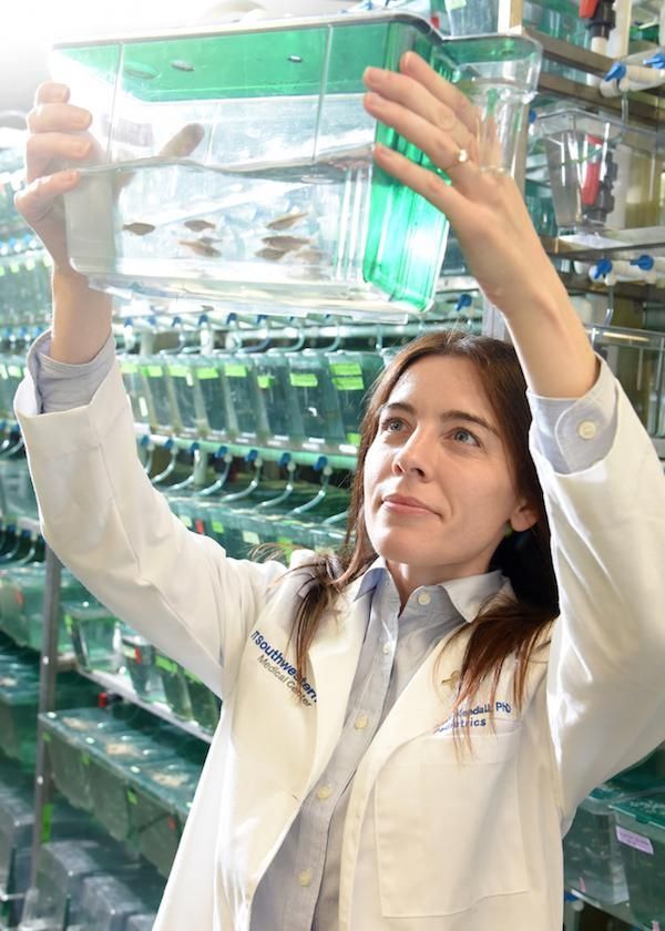
A popular aquarium fish may hold answers to how tumors form in a childhood cancer.
Muscle precursor cells called myoblasts are formed during normal fetal development and mature to become the skeletal muscles of the body. Rarely, a genetic error in which pieces of two chromosomes fuse together occurs in a cell related to this process and triggers those cells to multiply and behave abnormally. A particularly aggressive form of the muscle cancer rhabdomyosarcoma results.
The fused genes create an abnormal protein called PAX3-FOXO1, which blocks the normal maturation of muscle cells by inappropriately turning hundreds if not thousands of genes on and off. The exact mechanism by which PAX3-FOXO1 does this is not known.
Cancer researchers at UT Southwestern Medical Center developed a zebrafish model for the childhood cancer. To do this, Dr. James Amatruda’s lab inserted the human PAX3-FOXO1 gene into the DNA of zebrafish. Using this new transgenic zebrafish, the researchers showed that the fused-gene DNA causes rhabdomyosarcoma that is similar to the human disease. They found it does this by turning on another gene, HES3, which leads to overproduction of the skeletal muscle precursor cells and allows for PAX3-FOXO1+ cells to survive during development instead of dying.
Read more at UT Southwestern Medical Center
Image: Dr. Genevieve Kendall came to UT Southwestern because she wanted to work with zebrafish, which are an excellent model for studying childhood cancer. Because the young fish develop outside the mother's body, it's easy to insert human cancer-related genes into the fish genome, and drugs can be tested simply by adding them to the water. (Credit: UT Southwestern)


