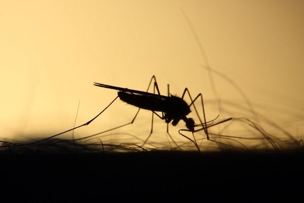
They approach with the telltale sign – a high-pitched whine. It’s a warning that you are a mosquito’s next meal. But that mosquito might carry a virus, and now the virus is in you. Now, with the help of state-of-the-art technology, researchers at the University of Missouri can see how a virus moves within a mosquito’s body, which could lead to the prevention of mosquitoes transmitting diseases.
“Previously, the common understanding was that when a mosquito has picked up a virus, it first needs some time to build up inside the midgut, or stomach, before infecting other tissues in the mosquito,” said Alexander Franz, an assistant professor in the Department of Veterinary Pathobiology in the MU College of Veterinary Medicine and the study’s corresponding author. “However, our observations show that this process occurs at a much faster pace; in fact, there is only a narrow window of 32 to 48 hours between the initial infection and the virus leaving the mosquito’s stomach. For this field of research, that revelation is eye opening.”
In this study, Franz and the team of researchers observed a mosquito infected with the chikungunya virus, which originates in Africa and was first found in the Americas in 2013. There is no vaccine to prevent or treat this virus, and while most common symptoms include fever and joint pain, they can be severe and disabling. The researchers used three separate electron microscopes to view the virus traveling through the mosquito, beginning with its midgut, or stomach. The first two microscopes provided different two-dimensional views of a single layer of tissue in the mosquito’s stomach. The third, a focused ion beam electron microscope, allowed researchers to see multiple layers of tissue.
Read more at University of Missouri-Columbia
Photo Credit: Emphyrio via Pixabay


