Biophysicists from the Moscow Institute of Physics and Technology have joined forces with colleagues from France and Germany to create a new fluorescent protein. Besides glowing when irradiated with ultraviolet and blue light, it is exceedingly small and stable under high temperatures. The authors of the paper, published in the journal Photochemical & Photobiological Sciences, believe the protein holds prospects for fluorescence microscopy. This technique is used in research on cancer, infectious diseases, and organ development, among other things.
Fluorescence microscopy is a method for studying living tissue that relies on induced luminescence. After being exposed to laser radiation at a particular wavelength, some proteins emit light at a different wavelength. This induced “glow” can be analyzed using a special microscope. Researchers append such fluorescent proteins to other proteins via genetic engineering to make the latter ones visible to the microscope and observe their behavior in cells. Fluorescence microscopy proved so scientifically valuable that one Nobel Prize was awarded for its discovery, followed by another one for radically improving the method’s accuracy.
Read more at Moscow Institute of Physics and Technology
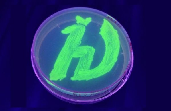
Photo: Petri dish with bacteria genetically modified to produce a fluorescent protein. The glowing symbol is a logo of the Moscow Institute of Physics and Technology. Image courtesy of the researchers.


