Electron microscopy is built on a bitter irony: The process of beaming electrons allows scientists to peer into the atomic structure of biomolecules and materials, but the electrons can also damage their target.
“I can only deliver so many electrons before I fry the sample, but I need a certain number of electrons before I can see the things that I need to see,” said David Muller, the Samuel B. Eckert Professor of Engineering in the Department of Applied and Engineering Physics. “Otherwise, everything’s just lost in this speckly noise. So there’s a trade-off.”
In 2017, Muller co-led a collaboration with Sol Gruner, the John L. Wetherill Professor of Physics, on a high-powered detector that, in combination with a technique called ptychography, set a world record for nanoscale resolution, measuring down to 0.39 ångströms or 0.039 nanometers; a nanometer is one-billionth of a meter.
Now, Muller’s research group has gone a step further by developing a new form of ptychography that uses complex algorithms to achieve faster, more efficient imaging with picometer (one-trillionth of a meter) precision that won’t damage samples so easily.
Read more at Cornell University
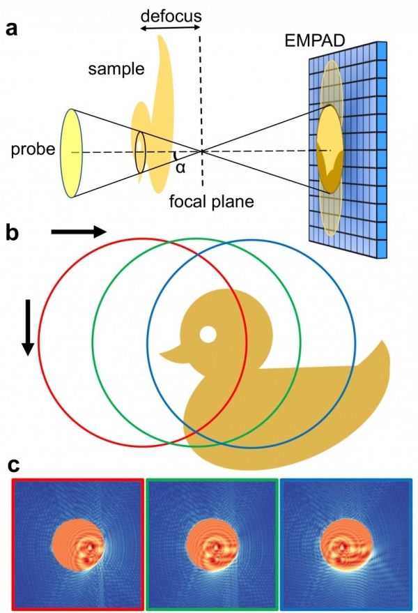
Image: This schematic shows how an electron probe is defocused to capture a wide range of data that is reconstructed into an ultraprecise image. The bottom three images are the diffraction patterns simulated when the probe is illuminated at the positions circled above. CREDIT: Cornell University